Table of Contents
-
Class Heterotricha, Subclass Heterotrichea (Blepharisma, Climacostomum , Condylostomum, Spirostomum and Stentor )
-
Ciliates Belonging To The Class Spirotricha, Subclass Hypotrichia (Aspidisca and Euplotes)
-
Class Spirotricha and Subclass Oligotricha: Halteria And Strombidium
-
Class Spirotricha, Subclass Stichotrichia (Paruroleptus Stylonychia , and Tachysoma)
-
Class Litostomatea,Subclass Trichostomatia (Lacrymaria, Litonotus,Phialina, Spathidium and Trachelius)
-
Class Nassophorea: Nassula
-
Class Prostomatea: Coleps
-
Class Oligohymenophorea, Subclass Peniculia- Lembadion and Paramecium
-
Class Oligohymenophorea, Subclass Scuticociliata (Cohnilembus, Cyclidium and Pleuronema)
-
Class Oligohymenophorea, Subclass Hymenostomata-Colpidium
-
The Ciliate Class Oligohymenophorea,Subclass Peritrichia- Campanella, Ophryidium, Vorticella, Trichodina and Zoothamnion
Protozoans, are generally microscopic, unicellular organisms that are classified by the cellular structures responsible for movement (Pseudopodia, Flagella and Cilia). Protozoans possess membrane bound cellular organelles such as Nuclei, Food Vacuoles, and Lysosomes. Food is captured in many different ways, however in most cases, after capture, it is contained in a spherical bubble (Food Vacuole ) that is formed from the cell membrane and released into the cell interior.Food vacuoles fuse with membrane bound lysosomes that contain digestive enzymes.Enzymes break down food into units that the protozoans can use for their own metabolic needs. In freshwater environments, water constantly passes into protozoan cells osmotically. If unchecked, the cell membrane will stretch and rupture. To counter this process most ciliates exposed to fresh or water of low salinity have contractile vacuoles that collect the incoming water and then move it to the outside.
1. Ciliate Genera Belonging To The Class Heterotricha, Subclass Heterotrichea (Blepharisma, Climacostomum , Condylostomum, Spirostomum and Stentor )
a. Bleparisma
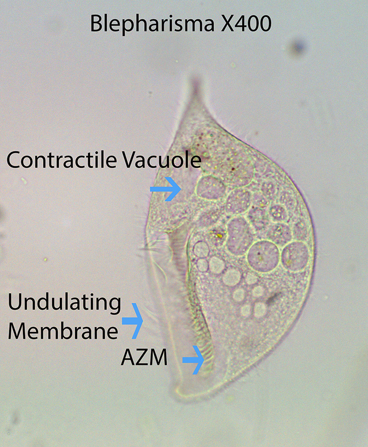
Bleparisma, one of the largest ciliates, is about 250 microns long. The elliptical body is narrower anteriorly and laterally compressed. Cilia associated with the mouth (Adoral Zone of Membranelles (AZM) extends posteriorly to about one-third of the body length. A transparent Undulating Membrane made up of long cilia fused together to form a single sheet, lies on the opposite side (Right) of the Adoral Zone of Membranelles (AZM). Both of the above mentioned structures move particulate food along a groove towards the mouth (Cytostome) where it is ingested. Many of the food vacuoles inside the cytoplasm are filled with bacteria. The cell body is uniformly ciliated (Somatic Cilia). Some of the cells are pale pink to light red. A Contractile Vacuole is situated near the posterior end. The feeding process is like that described for Ophrydium and Paramecium.
http://vimeo.com/115012399 (Zottoli) Blepharisma X100 Type 1. Movement
http://vimeo.com/115012400 (Zottoli) Blepharisma
X400 X1000 Type 1. At the beginning of the video, bacteria and small flagellates can be seen moving around as they are forced into a newly formed food vacuole. The AZM , Undulating Membrane , numerous Food Vacuoles, and a posterior Contractile Vacuole are visible.
http://vimeo.com/114933753 (Zottoli) Blepharisma
X400. Type 1. Anatomy
http://vimeo.com/114933746 (Zottoli) Blepharisma
X400 Type 2. The anatomical features are similar to those described in Type 1.
http://vimeo.com/114933749 (Zottoli) X400 X600. Type 2. Anatomy
https://vimeo.com/124156314 (Zottoli) X400 (Best)
b. Climacostomum
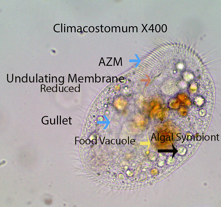
Climacostomum, about 150 microns long, is uniformly covered by somatic cilia. An Adoral Zone of Membranelles (AZM) curves around the front of the cell from left to right. A transparent Undulating Membrane made up of long cilia fused together to form a single sheet, lies on the opposite side (Right) of the Adoral Zone of Membranelles (AZM). Both help direct food towards the mouth. The AZM also is responsible for movement.The cell body is somewhat oval with blunt anterior and posterior ends. A few pale green endosymbiotic algae are visible in the cytoplasm. Food vacuoles often contain diatoms.
http://vimeo.com/115014656 (Zottoli), X400. Movement, Anatomy
http://vimeo.com/115016277 (Zottoli) X400. Movement and Anatomy
http://vimeo.com/115016275 (Zottoli) X400. Movement and Anatomy
c. Condylostoma
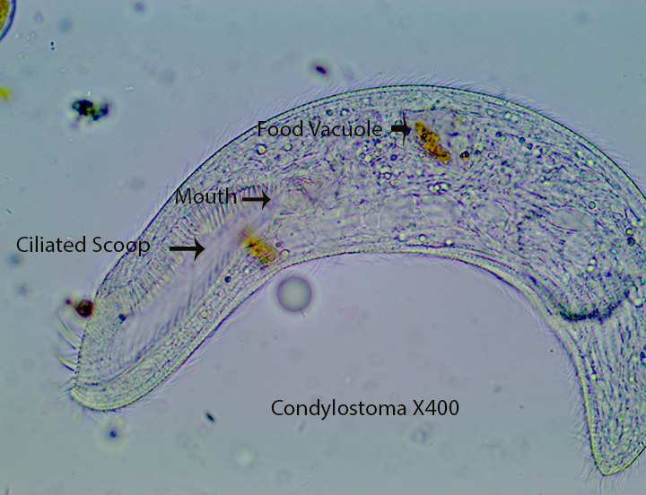
Condylostoma, also one of the largest ciliates, is about 400 microns long. The elongate extremely flexible cell body is uniformly ciliated (Somatic Cilia). An Adoral Zone of Membranelles (AZM) curves around the front of the cell from left to right. A large Undulating Membrane made up of long cilia fused together to form a single transparent sheet lies on the opposite side (Right) of the AZM (Visible). Both of these structures create water movement towards the mouth located just posterior to the end of the AZM. Condylostoma feeds on large and small diatoms as well as algae and protozoans.
http://vimeo.com/115065788 (Zottoli) X100 X400. Movement at 100X and Anatomy at 400X. Large, elongate diatoms as well as some type of green alga consumed by the ciliate, is evident inside the cell. The AZM is also visible.
http://vimeo.com/115065792 (Zottoli) X400. As above
http://vimeo.com/115065791 (Zottoli) X400. As above.
http://vimeo.com/115065790 (Zottoli). X400. Three large, elongate diatoms and two smaller ones are visible inside the cell.
d. Spirostomum
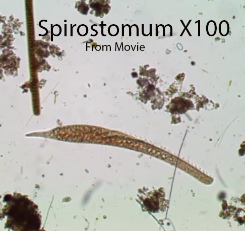
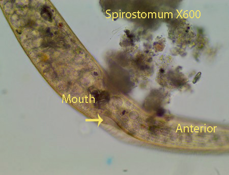
Spirostomum is a long, thin ciliate (Up to 1 mm in length) with a distinct, large, round, clear contractile vacuole at the posterior end. The anterior end of the cylindrical ciliate is narrower than the posterior. The uniformly ciliated cell body is extremely flexible as it navigates over and under obstacles in its path. The animal can move smoothly in a straight line as well as twist and turn as it navigates through debris. Often it will move forward a short distance and then backward and forward again. An adoral zone of membranelles (AZM) leads from the anterior end to the cytostome (Cell Mouth) located where the narrow anterior abruptly widens (About 1/3 of the way towards the posterior end). The body is uniformly ciliated and rows of cilia are visible. The macro-nucleus, visible in some of the specimens, looks like a “string of sausages.” It appears to feed on detritus, bacteria and small protozoans
http://vimeo.com/115067437 (Zottoli). X100X400. Movement and Anatomy. The bean-shaped structure inside the cell appears to be part of the macronucleus
http://vimeo.com/115067438 (Zottoli) X400. Anatomy.
http://vimeo.cm/115067439o (Zottoli). Anatomy.X400
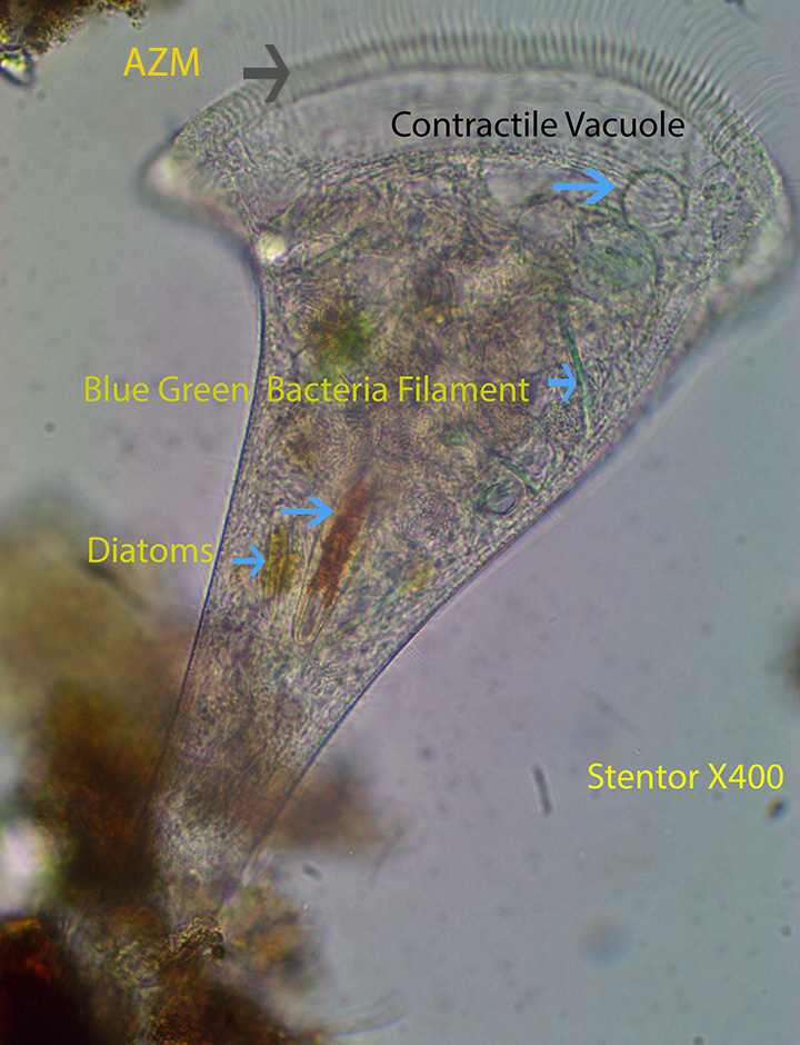
e. Stentor

Stentor, about 1 mm long, is characterized by its trumpet-shaped body. The anterior end is the widest and bears an adoral zone of membranelles (AZM) which create water currents that direct food towards the mouth. The posterior, thinner end (Holdfast) can attach to the substratum holding the animal in place. The ciliate can detach the holdfast and move to a new location. There also are cilia (Somatic Cilia) covering the entire cell body. The Contractile Vacuole is situated as shown above at the anterior end.
http://vimeo.com/115068793 (Zottoli) Diatoms, Bluegreen bacterial filaments, and tiny round bacteria are incorporated into food vacuoles internally
http://vimeo.com/115070092 (Zottoli). As above
2. Ciliates Belonging To The Class Spirotricha, Subclass Hypotrichia (Aspidisca and Euplotes)
a. Aspidisca
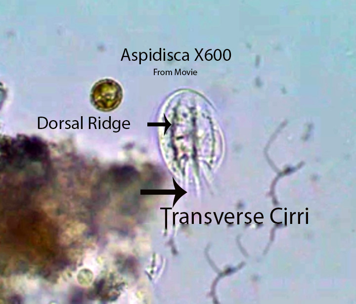
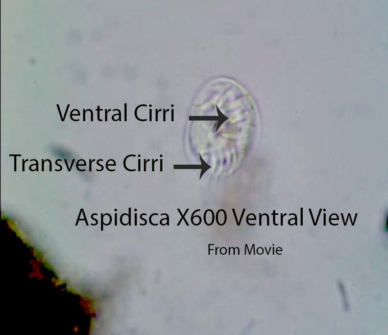
Aspidisca, about 50 microns long, is one of the most abundant ciliates in almost all of the samples collected from April to September. They literally “walk” over the substratum using the fronto-ventral (labeled Ventral) and transverse cirri. The fronto-ventral cirri are more active than transverse cirri and seem to be mainly responsible for movement. They move over clumps of detritus, then stop for a short time, twitch, and move again. Movement appears to be random. The cell body is inflexible and has four longitudinal dorsal ridges that extend from anterior to posterior. They have an adoral zone of membranelles (AZM) mounted on a stalk (not visible). The AZM is moved over the substratum collecting bacteria, detritus, small protozoans, etc. Bacteria appear to be its main source of food.
http://vimeo.com/114829411 (Zottoli) X400. Movement and Anatomy
http://vimeo.com/114829410 (Zottoli) X400. Movement and Anatomy
b. Euplotes
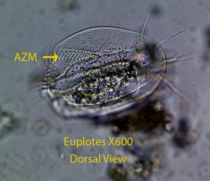
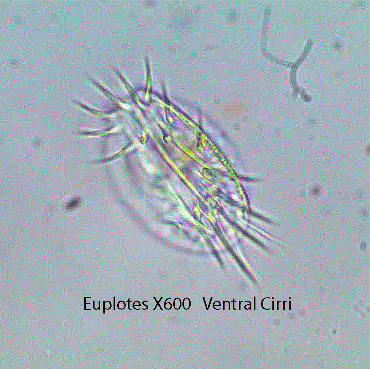
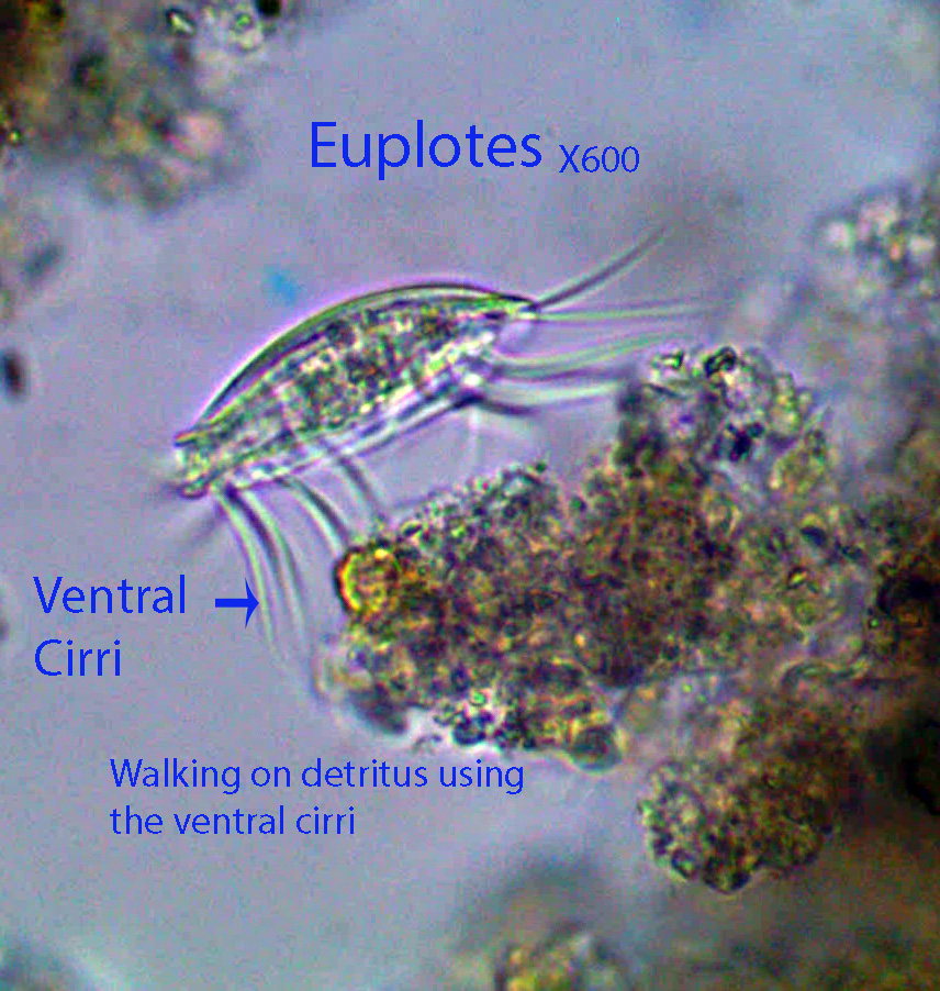
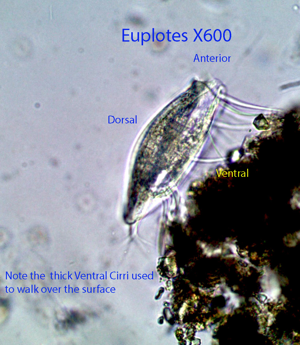
Euplotes, about 50 microns long, has an adoral zone of membranelles (AZM) ,with long cilia that pull water beneath the cell towards the mouth. Suspended particles are removed from the water and pass into the cell interior. They feed on detritus, bacteria, small protozoans, etc. Water circulation is visible in some of the videos. The ventral surface of the cell is flattened and the dorsal surface is rounded (Convex). . The AZM also moves the rigid cell body forward when they travel through the water column. Euplotes has 4 frontal cirri that may have a sensory function and 9 thick ventral cirri, visible in the videos, that are used to walk on the substratum. 4-6 posterior, caudal cirri extend posteriorly from the ventral surface and 5 anal cirri are located at the posterior end; both of these groups seem to stabilize (balance) the cell body during movement.
http://vimeo.com/114831678 (Zottoli) X400 X600. Ventral view. The ventral cirri are pointed upward, touching the bottom of the microscopic cover glass. The use of individual ventral cirri can be followed as the animal moves forward.
http://vimeo.com/114876795 (Zottoli) X600.Water circulation is clearly shown.
http://vimeo.com/114876794 (Zottoli) X400. Movement
http://vimeo.com/114879023 (Zottoli) X600. Anatomy
3. Ciliates Belonging To The Class Spirotricha and Subclass Oligotricha: Halteria And Strombidium
a. Halteria
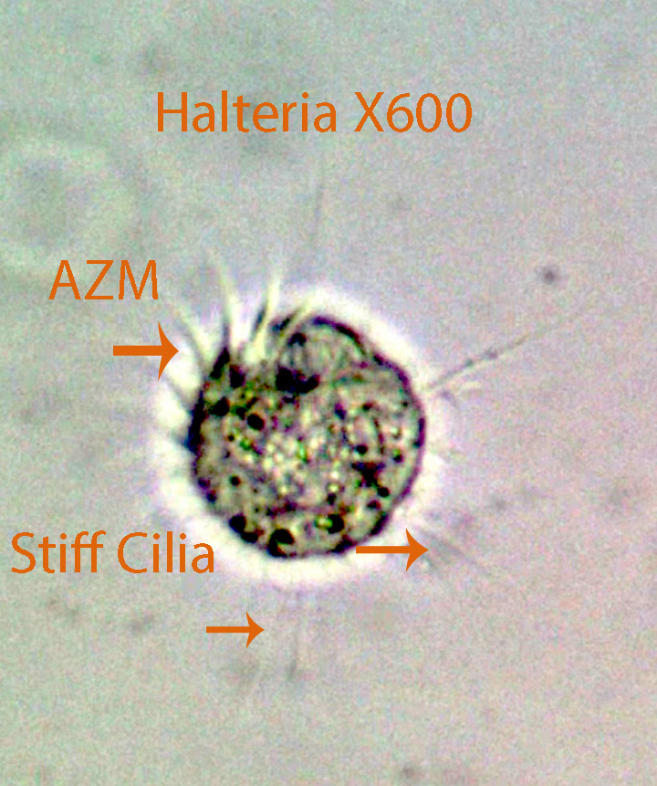
Halteria, about 30 microns long, is one of the most common ciliates here. It literally hops from place to place randomly. There is a circle of cilia (Prominent AZM) around the mouth at shown above. Locomotion is caused by the quick movement of stiff cilia around the equator of the cell. It appears as if they feed when they stop moving. The anterior cilia (AZM) create water movement that moves particulate matter towards the mouth.
http://vimeo.com/114375614 (Zottoli) X400
http://vimeo.com/116179643 (Zottoli) X600
http://vimeo.com/114375615 (Zottoli) X600
b. Strombidium
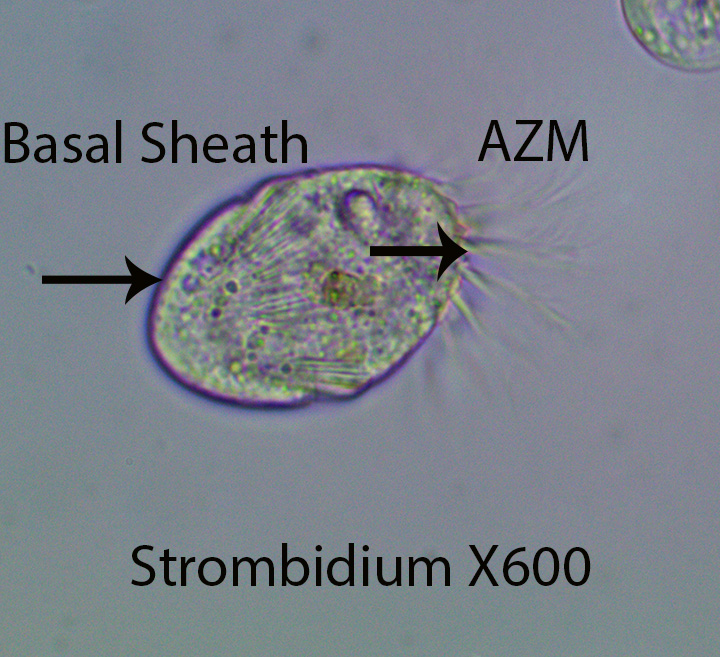
Strombidium, about 30 microns long, has a strongly developed adoral zone of membranelles (AZM) around the anterior end that is responsible for movement and feeding. When the cilia beat, the cell moves forward, swaying slightly from side to side. There is a basal sheath (lorica) around the posterior part of the cell. One contractile vacuole lies near the cell center. The ciliate feeds on bacteria, diatoms and small, round, green algal cells.
https://vimeo.com/120887419 (Zottoli) (Strombidium)
4. Ciliates Belonging To The Class Spirotricha, Subclass Stichotrichia (Paruroleptus Stylonychia , and Tachysoma)
a. Paruroleptus
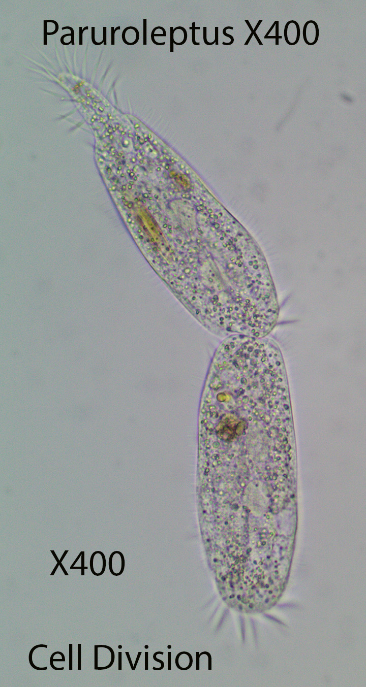
Paruroleptus, about 350 microns long, has two straight rows of marginal (along the edge) cirri as well as two rows of mid-ventral cirri arranged in a zig-zag pattern. The flexible ciliate has an adoral zone of membranelles (AZM) that create water flow towards the mouth. After entering the mouth food particles are directed down to the base of the cytopharynx where a food vacuole is formed. They feed on bacteria, diatoms and small protozoans. The cell body narrows at the posterior end forming a sort of tail.
http://vimeo.com/114887341(Zottoli) X400. Movement. Note the presence of diatoms inside the ciliate.
http://vimeo.com/114891224 (Zottoli) X400. Movement and Anatomy
http://vimeo.com/114891223 (Zottoli) X400. Movement and Anatomy
http://vimeo.com/114892990 (Zottoli) X400. The stationary picture that you see before starting the video clearly shows the following: The clear round Contractile Vacuole; The rounded two part Macronucleus (more dense than the contractile vacuole) and two Food Vacuoles filled with a green alga of some type
http://vimeo.com/114892991 (Zottoli) X600. Movement and Anatomy
b. Stylonychia
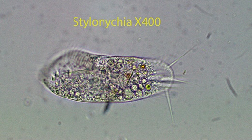
Stylonychia, about 160 microns long, has a rigid, elliptical cell body with 3 (2 lateral and one central), obvious caudal cirri. A prominent AZM (Adoral Zone of Membranelles) composed of many cilia, forms a “collar” from right to left around the front of the cell body. They aid in procuring food and in locomotion.There are two rows of marginal (along the edges) cirri, however they do not continue around the posterior end. The marginal cirri aid in movement. Groups of cirri on the ventral surface allow the ciliate to “walk” on solid surfaces and also move quickly from place to place. There is an undulating membrane on the right side, across from the AZM. They feed on a variety of small organisms such as bacteria, protozoans, algae, etc. and also on large prey such as diatoms. A Contractile Vacuole is located internally near the posterior end of the AZM.

http://vimeo.com/114899505 (Zottoli)X400. Stylonychia is in the final stage of dividing into two identical individuals. The AZM is visible as are several greenish food vacuoles . Notice how the cilia in the AZM move
http://vimeo.com/114901510 (Zottoli) X400. General Characteristics. Movement appears to be random and jerky
http://vimeo.com/114901509 (Zottoli) X400. General Characteristics. The two rows of marginal (Lateral) cirri are visible along with the AZM (Adoral Zone of Membranelles). In the latter part of the video I focused downward to show the ventral cirri
c. Tachysoma
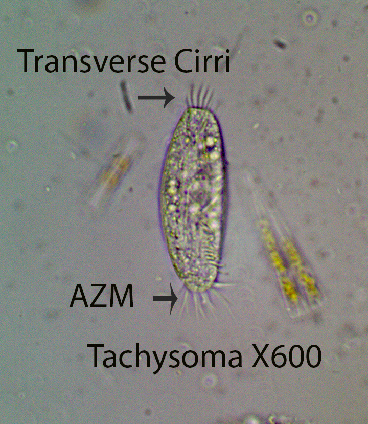
Tachysoma, about 100 microns long, has a relatively rigid cell body with long, dorsal, thin, immobile Bristles. A small AZM forms a “collar” from left to right around the anterior of the cell body. Two marginal (Along the edges) rows of cirri are present that do not extend around the posterior end. Five transverse cirri are situated at the posterior end. A contractile vacuole is located at about the center of the cell. All three videos show the AZM, the 5 transverse cirri and the 2 marginal (Lateral) rows of cirri.
https://vimeo.com/149443613 Tachysoma X600 Movement on Enteromorpha filament
http://vimeo.com/114932454 (Zottoli) X400
http://vimeo.com/114932453 (Zottoli) X400. The photograph that appears before starting the video shows the following structures: The 5 Transverse Cirri at the posterior end; The Small AZM at the anterior end and one clear, round Contractile Vacuole on the right hand edge inside the cell body
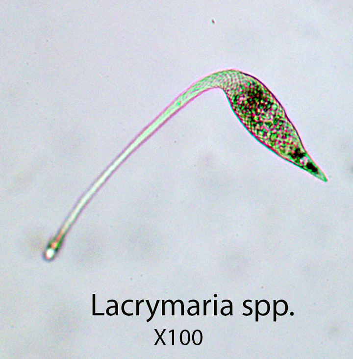
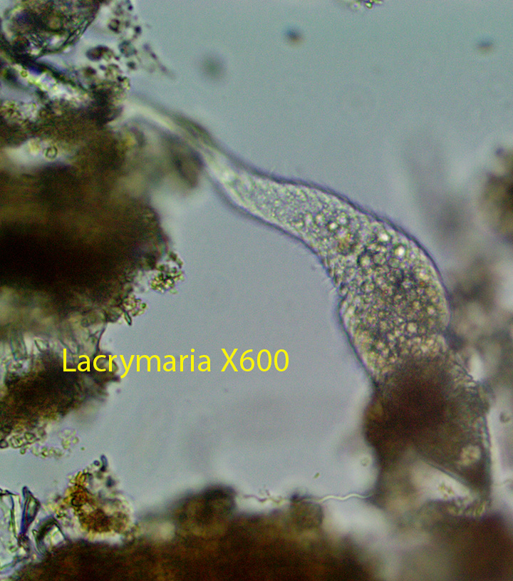
Lacrymaria, about 450 microns long, has a sac-like body with an extensible neck. The fusiform body makes it easier for them to move into and between clumps of detritus. The mouth (Cytostome) is situated at the tip of the neck. Organelles called extrusomes, located here,release a toxin that kills or quiets prey making them easier to ingest. The ciliate spends most of its time embedded in debris moving its neck back and forth in search of food. The neck can be extended to about twice the length of the body. Somatic cilia are arranged in spiral rows that are most evident on the posterior part of the cell body. They feed for the most part on small protozoans.
http://vimeo.com/114159113(Zottoli)X100
http://vimeo.com/114159115 Lacrymaria (Zottoli) X400
http://vimeo.com/114159114 Lacrymaria (Zottoli) X400
b. Litonotus
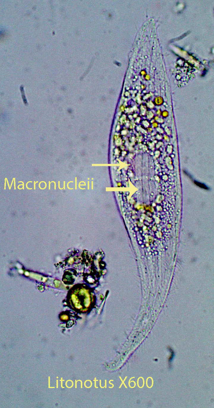
Litonotus, about 400 microns long, has a sac-like body with a relatively short, narrow neck. The somatic cilia are arranged in parallel rows. The neck is not as flexible or extensible as that in Lacrymaria. The mouth is located on the anterior convex, lateral surface and is equipped with extrusomes that produce a substance toxic to prey. The ciliate often moves forward and then backward when they encounter a clump of detritus. The anterior end probes the area around them. Two round macronucleii are visible inside the cell. A single contractile vacuole is situated at the posterior end. Litonotus is an active predator known to feed on other protozoans. The second video shows Litonotus feeding on a much larger ciliate (Euplotes). Round green algal cells , one per vacuole also have also been observed.
https://vimeo.com/148640615Litonotus X600
https://vimeo.com/150289604 Litonotus feeding on Euplotes
c. Phialina
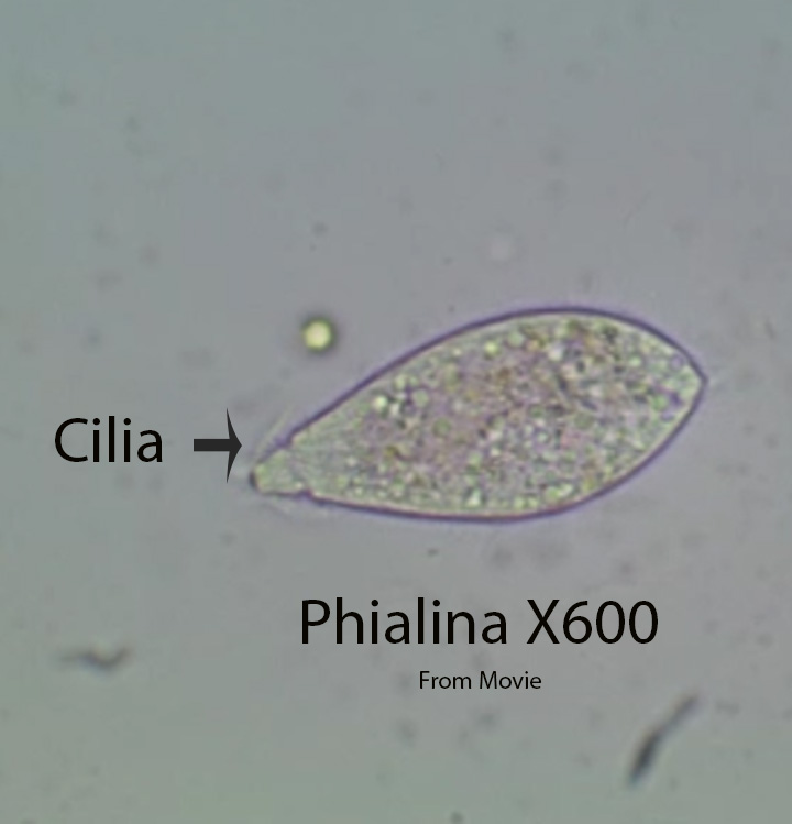
Phialina , about 80 microns long, is closely related to Lacrymaria based on its possession of a “short” neck with a mouth at its tip , extrusomes associated with the round mouth, and the presence in this region of a wreath of relatively long cilia just below the cell tip. Extrusomes are cell organelles that release secretions that immobilize prey. The neck however is not as long or as active as that found on Lacrymaria. A contractile vacuole is situated at the posterior end and a single macro-nucleus is present. Rows of cilia spiral around the cell body. Phialina moves quickly through debris, moving its short neck from side to side in what appears to be a search pattern. After a time, the animal may back out and move to a new location. Movement appears to be random. It can move forward, backward and bend from side to side.They feed for the most part on small protozoans.
http://vimeo.com/114249822 (Zottoli)X400
d. Spathidium
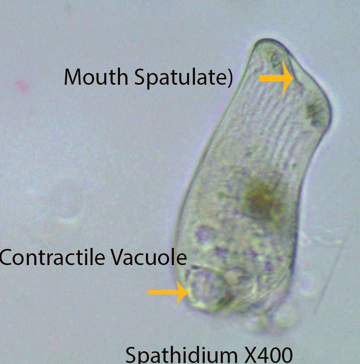
Spathidium is a sac-shaped, flattened ciliate about 150 microns long. The cell mouth is relatively wide (Spatulate) and bears a ring of cilia around its edge. Oral cilia are responsible for moving the body as well as directing food into the mouth. They also rotate around their anterior-posterior axis as they move forward. Extrusomes located around the oral opening help to kill and trap prey making it easier for the ciliate to ingest them. Spathidium is a predator that feeds on medium-sized protozoans and rotifers.
https://vimeo.com/124156315 (Zottoli) X100 X400
https://vimeo.com/124156313 (Zottoli) X400
e. Trachelius
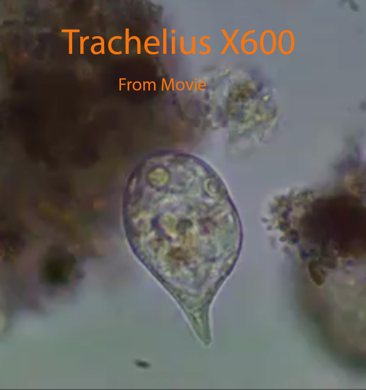
Trachelius, about 150 microns long, is distinguished by a short tapering neck and a round cell body. The round cell mouth (Cytostome) leads into a short tube (Cytopharynx) that ends internally. Food vacuoles are formed at the end of the cytopharynx. The Y-shaped structure (mouth and cytopharynx) are visible in the video below:
https://vimeo.com/81210133 (Trachelius X400) collected in a local bog.
The animal rotates around its central anterior-posterior longitudinal axis. Feeding was not observed, however detritus, bacteria and small protozoans were observed in food vacuoles.
http://vimeo.com/114348067 (Zottoli)X100
http://vimeo.com/114348068 (Zottoli) X400
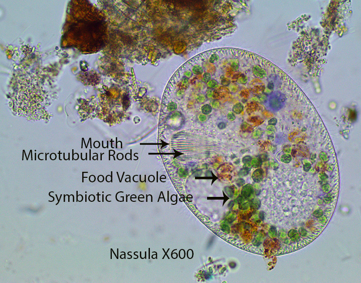
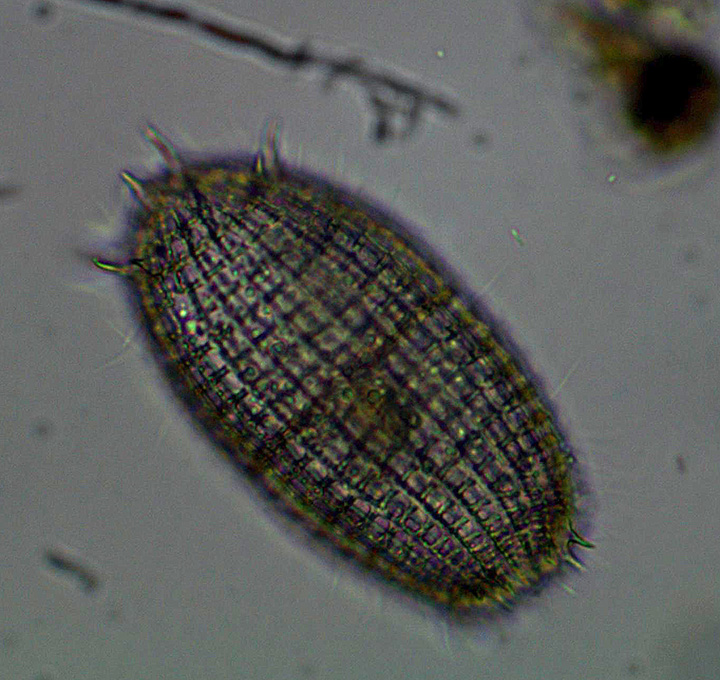
Nassula, about 200 microns long is dorso-ventrally flattened and uniformly ciliated. Some of the specimens harbor a large number of what appear to symbiotic green algae.. The ventral mouth, located near the anterior end, is surrounded and supported by a cylinder of micro-tubular rods that help draw in filamentous blue green bacteria such as Oscillatoria. In the video, Nassula (https://vimeo.com/149409555 ) grabs a straight Oscillatoria filament, bends it in half, and draws it smoothly into the cell. As blue green algae are digested the pigments change color from blue green to red, purple and orange. The ciliate moves in a more or less straight line until it comes in contact with a clump of detritus. At this point Nassula often pushes its anterior into debris presumably searching for food. They occasionally rotate around the central anterior-posterior longitudinal axis as they move forward.
7. Ciliates Belonging To The Class Prostomatea: Coleps
Coleps
Coleps, about 80 microns long, is covered by external plates secreted by the cell. The barrel-shaped cell body is uniformly ciliated; Cilia protrude through the calcium carbonate plates along visible longitudinal lines. Small spikes surround the anterior and posterior poles. A group of cilia surround the mouth. As the ciliates move forward they rotate from left to right or the opposite way, around their central anterior-posterior longitudinal axis. They also can move forward and backward without rotating. They often push their anterior end into chunks of debris containing bacteria and small algal cells moving slightly from side to side. Oral cilia move dislodged material into the mouth. Micro-tubular rods (not seen) form a circular channel from just below the mouth into the cell interior. They also are attracted to and feed on dead and dying organisms. A single contractile vacuole lies near the posterior end. Coleps is abundant throughout spring and summer. Food vacuoles may contain detritus, bacteria , small algal cells and diatoms.
http://vimeo.com/114255117 (Zottoli) X400. Coleps feeding on the Rotifer Notommata spp.
8. Ciliates Belonging To The Class Oligohymenophorea, Subclass Peniculia- Lembadion and Paramecium
a. Lembadion
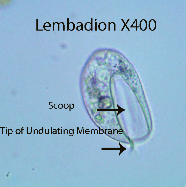
The predatory ciliate Lembadion, about 95 microns long, is characterized by its large, scoop-shaped oral cavity. There is an undulating membrane within the oral cavity that moves back and forth. The cell body rotates around the anterior-posterior axis as the animal moves forward. The tip of the undulating membrane extends slightly from the front of the scoop. The scoop might help the ciliate hold prey in place as they are forced into a food vacuole. Small flagellates and bacteria have been observed inside food vacuoles. The cell body is uniformly ciliated.
http://vimeo.com/114362966 (Zottoli) X1000. Although this specimen was collected from a Raised Bog, it is included because it is much larger than the species found here, making it easier to view anatomical features.
http://vimeo.com/114359947 (Zottoli) X400 X600. General Movement
http://vimeo.com/114363916 (Zottoli) X400. General Movement and Gross Anatomy
b.Paramecium
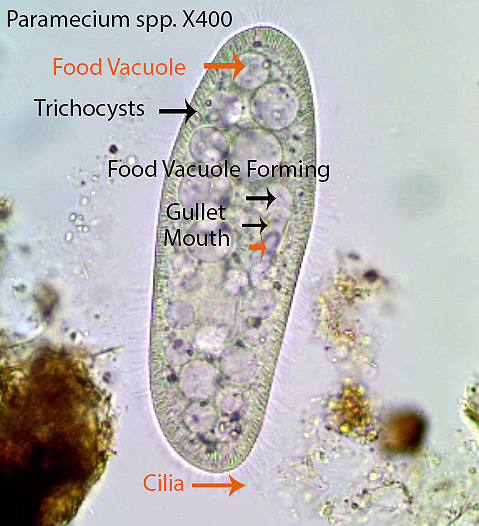
Paramecium is about 230 microns long. Trichocysts are visible as short vertical lines just beneath the cell membrane. When the ciliate is disturbed, a long thin, sticky thread is released from some or most of the trichocysts. Mass release of these filaments makes it difficult for protozoan predators to ingest them. This defensive function has recently been confirmed experimentally. A small number of threads, released from the same area, allow the ciliate to temporarily attach to the substratum, an advantage in food rich locations. A pre-oral groove, visible in some of the videos, leads from the anterior end to the mouth. The mouth is a circular opening that leads to a short tube (Gullet or Cytopharynx) that empties into a food vacuole. The vacuole fills with bacteria moved by cilia along the pre-oral groove and cytopharynx. The food vacuole eventually breaks away and moves into the cytoplasm. Food vacuole formation can be observed at the beginning of the first video:
http://vimeo.com/116576162 (Zottoli) X600.
In the video, the anterior end is towards the left. Food vacuole formation is visible about 1/3 of the way towards the anterior end near the bottom of the cell. Bacteria are circling inside the vacuole, driven by cilia in the gullet. A new food vacuole forms as soon as the old one leaves. Cell organelles called lysosomes, filled with digestive enzymes, fuse with food vacuoles, dumping their load into the vacuole interior, initiating the digestive process. After digestion is completed the depleted food vacuole merges with the anal pore and its contents are released outside the cell as shown in the following video:
http://vimeo.com/82801689 (Zottoli) X400
Cytoplasm moves inside the cell (Cyclosis) as shown in several of the videos. Paramecium living in freshwater take in water osmotically since the concentration of dissolved substances is greater inside the cell than outside. Organelles called contractile vacuoles, one at each end of the cell, receive excess water from radiating canals and squirt it outside the cell when they contract. This process can be observed in the following video:
http://vimeo.com/116576163 (Zottoli) X600.
https://vimeo.com/116576161 Paramecium X600
Additional Paramecium Videos:
http://vimeo.com/114357168 (Zottoli)X600.Anatomy
http://vimeo.com/114357165 (Zottoli) X600 Movement
http://vimeo.com/114354101 (Zottoli) X400. Endo Symbiosis, Cyclosis
http://vimeo.com/114161463 (Zottoli) X400.Cell Division
9. Genera Belonging to the Class Oligohymenophorea, Subclass Scuticociliata (Cohnilembus, Cyclidium and Pleuronema)
Scuticociliates are small, oval , relatively inflexible ciliates that filter feed for the most part on bacteria. The genera Cyclidium and Pleuronema, discussed below, are common components of the protozoan community.
a. Cohnilembus
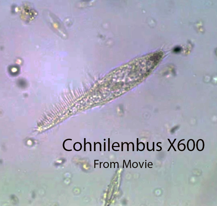
Conhnilembus, about 100 microns long, has a thin body that tapers anteriorly. The cell is uniformly covered by somatic cilia. Cilia at the posterior end are slightly longer than those on the cell body. Several, relatively long, bent cilia are present at the tip of the narrow anterior end. One row of buccal cilia lies on each side of an oral groove that leads to the mouth at about the middle of the body where the mouth (cytosome) and cytopharynx are located. A long, transparent undulating membrane,made up of long cilia fused together to form a single transparent sheet.extends from the anterior end to the middle of the body. A line of curved cilia lie alongside the undulating membrane. It often moves forward in a straight line, stops to feed and then moves again to a new location. It moves like Cyclidium, but not as fast. A contractile vacuole is located at the posterior end. They filter feed for the most part on bacteria.
http://vimeo.com/116174236 (Zottoli) X400
http://vimeo.com/116174237 (Zottoli) X400
b. Cyclidium
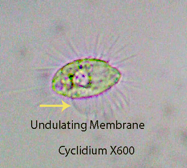
Cyclidium, about 20 microns long, is one of the most common ciliates living in the freshwater intertidal. The cells are oval in shape and relatively inflexible. They jump to a new site, feed for a short time and then quickly move again. The cell body is evenly ciliated except for a slight depression towards the anterior end where a transparent sheet-like undulating membrane (Visible) extends above the cell surface during feeding bouts. The somatic cilia stand straight out from the cell surface when the animal stops to feed. They consume bacteria for the most part.
Cyclidium, about 20 microns long, is characterized by the presence of relatively long cilia that extend outward at a 90 degree angle from the cell surface. This is most evident when the ciliate is still, during the feeding process. The cell body is evenly ciliated except for a slight depression towards the anterior end where a transparent sheet-like undulating membrane (Visible). made up of long cilia fused together, is located.The sheet,extends above the cell surface during feeding bouts. The undulating motion of the membrane directs small particles (mostly bacteria) into the mouth where they are incorporated into food vacuoles. If Cyclidium is disturbed or stops feeding, they “dart” to a new location usually not far from where they started, and begin feeding again. A single relativelyt long cilium protrudes from the posterior end. Cyclidium consumes bacteria for the most part. Although the feeding process is visible in this genus it can be seen with greater clarity in the anatomically similar Pleuronema.
http://vimeo.com/116174235 (Zottoli) X1000 . The specimens (Cyclidium) shown here were collected from a raised bog and shown here because they are larger than freshwater intertidal specimens. The sheet-like undulating membrane (visible) is extended from the cell surface as they feed.
http://vimeo.com/116176713 X400 (Zottoli) X600
http://vimeo.com/114266888 X400 (Zottoli) X400
c. Pleuronema

Pleuronema , about 80 microns long, is a close relative of Cyclidium. The somatic cilia are relatively long and stand straight out from the cell surface while feeding. They remain motionless while they feed. A relatively large, transparent undulating membrane made up of long cilia fused together to form a single transparent sheet,is extended from the cell body during the feeding process.It circulates water containing food towards the mouth and into the cytopharynx and into a food vacuole. Usually the vacuole fills with bacteria and eventually pinches off at its attached end and enters the cytoplasm where digestion takes place.
The entire process is clearly visible in the video: https://vimeo.com/148737306 Pleuronema X600
They filter feed on a variety of small organisms such as bacteria, algae, small protozoans, etc. After they have finished feeding, the long cilia quickly move them (jump/hop) to a new spot where they unfurl the undulating membrane and feed again.
https://vimeo.com/148737297 Pleuronema X600
http://vimeo.com/114302262 (Zottoli) X400
10. Ciliates Belonging To The Class Oligohymenophorea, Subclass Hymenostomata-Colpidium
Colpidium
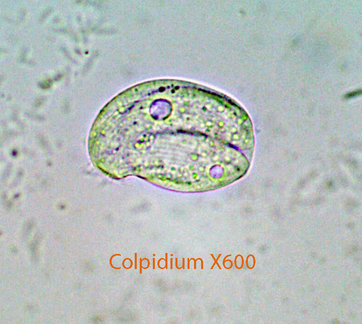
There is an indentation below the anterior part of the cell where the mouth is located. It is most evident in the first video where oral cilia create water currents that bring small particles towards the mouth. Bacteria seem to be the main source of food. The laterally flattened cell body, about 40 microns long, is rounded, pear-shaped, and uniformly ciliated. The front part of the cell is extended slightly to the side. A single contractile vacuole lies in the center of the cell. The cell body of most specimens is filled with food vacuoles.
http://vimeo.com/116391340 (Zottoli) X400. General Anatomy
11. The Ciliate Class Oligohymenophorea,Subclass Peritrichia- Campanella, Ophryidium, Vorticella, Trichodina and Zoothamnion
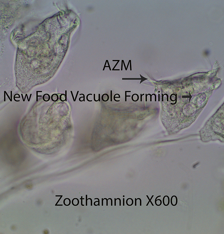
Peritrichs generally are bell-shaped ciliates that are attached to the substratum by stalks. In some species the stalk has a centrally located contractile thread that can pull the individual or colony downward quickly, out of harms way. They are characterized by an oral ciliated membrane that starts in the oral cavity and winds spirally in a counter clock wise direction to a point along the edge of the area that surrounds the mouth. There are no somatic cilia on the cell body.
1. Campanella
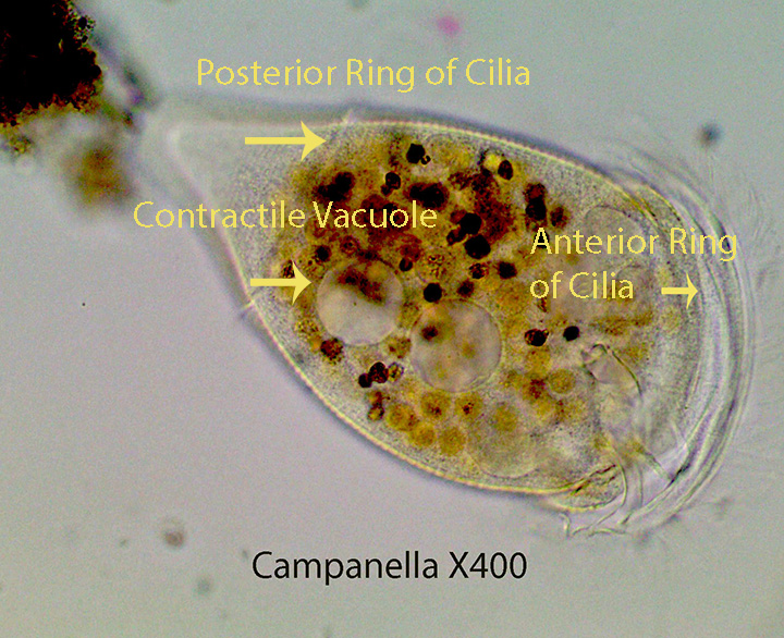
Campanella, one of the larger ciliates, is about 250 microns long. The anterior ring of cilia around the oral surface creates a vortex that drives particulate food towards the mouth. The food is passed from the mouth to the gullet and then into the rounded food vacuole (Visible at the beginning of the video). The feeding process is like that described for Ophrydium and Paramecium. It is normally attached by a stalk; however the specimen shown here was swimming, aided by the anterior ring of cilia around the mouth and the ring of posterior cilia that is present only in migrating cells (Telotroch Larva). They basically feed on bacteria, algae, and protozoans, etc.
http://vimeo.com/114365252 (Zottoli) X400
http://vimeo.com/114365251 (Zottoli) X400
http://vimeo.com/114365253 (Zottoli) X400
2. Ophrydium
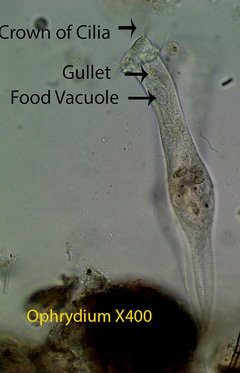
The ciliated protozoan Ophrydium, about 300 microns long, has a crown of cilia around its anterior end. The cilia create a current of water that directs particulate matter in the water column towards the “mouth (located in the center of the crown). Food then passes down a short gullet into a football-shaped food vacuole (Visible). The small dots inside food vacuoles are bacteria; however larger green flagellated protozoans are common prey. This process is called filter or particulate feeding. The single food vacuole breaks free and drops into the cytoplasm (Visible) and fuses with membrane bound lysosomes (Not Visible) that contain digestive enzymes. The video of Ophrydium feeding was filmed from specimens that I collected at a local bog. I included it here because it is exceptionally clear. The enzymes break down food into units that the protozoans can use for their own metabolic needs.
http://vimeo.com/114163634 (Zottoli) X400
3. Trichodina
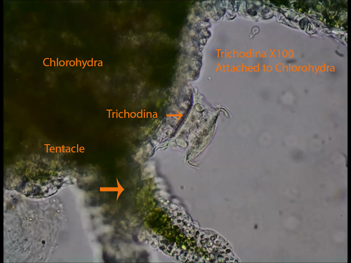
Trichodina lives as an ecto-commensal on the surface of Hydra (Phylum Cnidaria). The first part of the video shows the body and tentacles of Hydra. Trichodina appears towards at the beginning and at the end of the video; They are box-shaped and are attached to the outside of the tentacle. Oral cilia on the anterior (Upper) end are responsible for creating water movement towards the mouth allowing the animal to consume (Filter Feed) organisms such as bacteria and small protozoans as well as detritus. Posterior cirri grasp and pull the ciliate down on the epidermis of Hydra possibly creating suction that might help the ciliate maintain it’s position while feeding.
http://vimeo.com/114368245 (Zottoli) X100
4. Vorticella
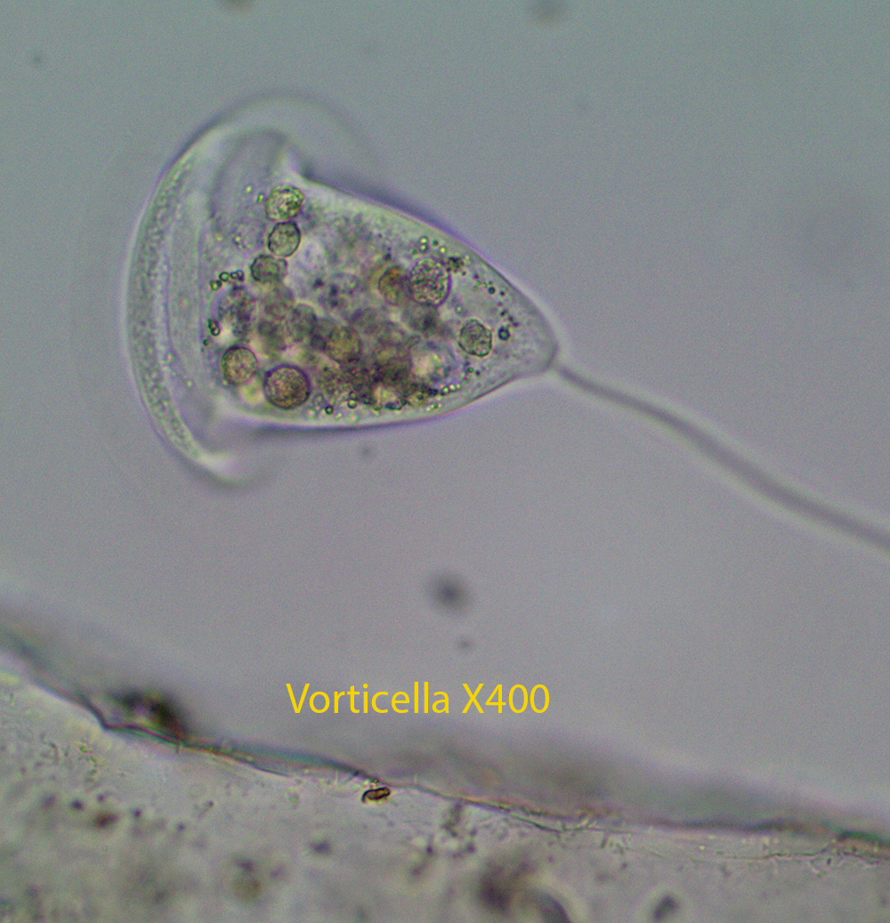
Vorticella is similar anatomically to Zoothamnion described below except that it is not colonial. One cell, about 50 microns long, is attached to one contractile stalk.

Leave a comment