Difflugia, about 120 microns long, is housed in a vase-shaped test covered with a few adherent quartz grains. There is a round opening (Aperture) circled by a lip at the anterior end through which the clear pseudopods extend. In one instance, two pseudopodia encircled a detrital mass. Part of the detrital mass containing bacteria and small protozoans was ingested at the base of the tentacles indicating that one role of the tentacles is to move detritus to the cell opening. During this process the pseudopods were clear. Small, round green algal cells (most likely symbiotic) were observed internally along the edge of the cell membrane.
Genera of Amoeboid Protozoans Commonly Found in the Freshwater Intertidal: Amoeba, Arcella, Centropyxis, Cyphoderia, Difflugia, Euglypha, Hartmanella ,Mayorella Saccamoeba, Thecamoeba and Trinema. Three Heliozoans are also described at the bottom of the page (Acanthocystis, Actinophrys and Actinospherium).
Introduction:
Protozoans, are generally microscopic, unicellular organisms that are classified by the cellular structures responsible for movement (Pseudopodia, Flagella and Cilia). Protozoans possess membrane bound cellular organelles such as Nuclei, Food Vacuoles, and Lysosomes. Food is captured in many different ways, however in most cases, after capture, it is contained in a spherical bubble (Food Vacuole ) that is formed from the cell membrane and released into the cell interior. Food vacuoles fuse with membrane bound lysosomes that contain digestive enzymes. The enzymes break down food into units that the protozoans can use for their own metabolic needs. In freshwater environments, water constantly passes into protozoan cells osmotically. If unchecked, the cell membrane will stretch and rupture. To counter this process most amoeboid protozoans exposed to fresh water or water of low salinity have contractile vacuoles that collect incoming water and then move it to the outside.
Amoeboid protozoans do not possess cilia and generally lack flagella. They may be protected by an external covering (Test) or they may lack such a covering (Naked) and therefore be directly exposed to the environment. The protective tests, secreted by the cell body may be constructed of organic material, silicon or a sticky matrix that particles such as small sand grains adhere to.
Types of Amoebas:
a. Naked amoebas, often form broad, finger-like extensions called Lobopodia or Pseudopodia:
(Lobopodia): http://vimeo.com/115444163 (Zottoli) (Amoeba) X400.
b. Test forming amoebas may also form lobopodia:
http://vimeo.com/115439340 (Zottoli) Difflugia X400)

c. Some form thin extensions called Filipodia:
http://vimeo.com/115670878 (Zottoli) Cyphoderia X400)
Descriptions of Amoeboid Genera
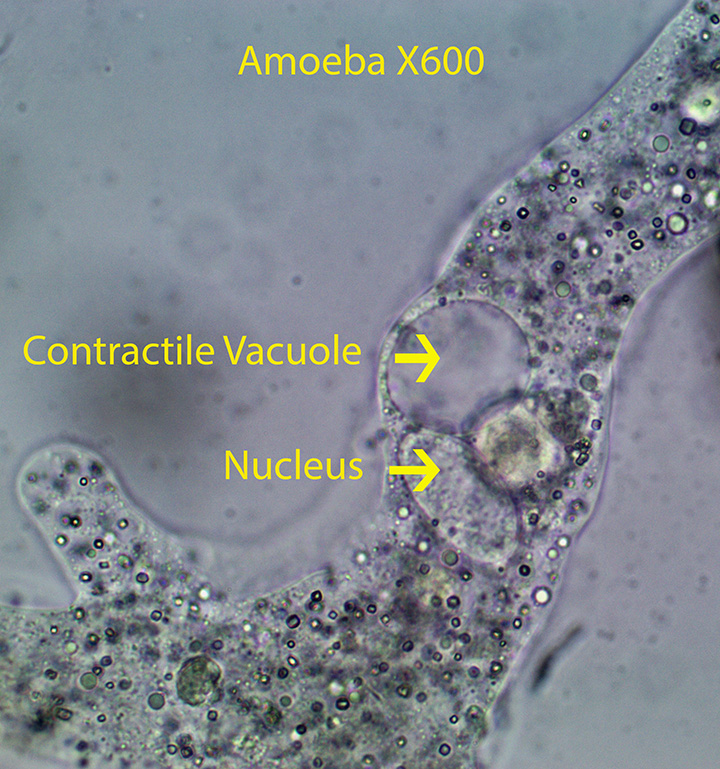
1. Amoeba (Amoebidae)

Amoeba feeds by enclosing bacteria or small protozoans such as flagellates in part of their cell membrane forming a circular food vacuole. Food is digested in the vacuole when it fuses with lysosomes that contain digestive enzymes. Certain amoeba species can reach one mm in length. The photograph (X600) above and the videos (X600 and X1000) shown below, showcase a naked (Lacks a Test) amoeba belonging to the genus Amoeba. They move by extending several blunt projections (Lobopodia). The central cytoplasm (Endoplasm) within the pseudopod flows anteriorly (Sol), pushing the cell forward. When it reaches the tip of the pseudopod, cytoplasm turns to Gel within the clear outside layer (Ectoplasm). Ectoplasm passes posteriorly where it eventually converts to Sol and moves forward again. This process was described at the molecular level relatively recently. A football shaped nucleus is clearly visible along with the perfectly round, clear contractile vacuole. The contractile vacuole (CV),is almost always visible inside the cell. It is responsible for passing water that enters the cell osmotically to the outside.
http://vimeo.com/115441424 (Zottoli) X600
http://vimeo.com/115441422 (Zottoli) X600
http://vimeo.com/115444160 (Zottoli) X600
http://vimeo.com/115722784 (Zottoli) X1000
http://vimeo.com/115439338 (Zottoli) X1000
2. Arcella (Arcellidae)

Arcella is covered by a circular, shell-like, textured covering (Test) as seen in the photograph above, with a ventral round opening through which clear, blunt pseudopodia (Lobopodia) extend to the outside. Pseudopods move the animal slowly forward as shown in the videos below. They are also responsible for moving detritus containing bacteria and small protozoans into the center of the ventral opening where it is incorporated into food vacuoles. Some contain symbiotic circular green algae internally. Often the algae are located on the outer edge of the cytoplasm. Thin cytoplasmic extensions attach the animal to the inside sides of the “shell” providing stability. In the last video note the relatively large rotifer trapped inside the “shell”. I suspect that the rotifer will ultimately end up in a food vacuole.
http://vimeo.com/115447167 (Zottoli) X400
http://vimeo.com/115444162 (Zottoli) X600
3. Centropyxis (Centropyxidae)

Centropyxis, about 160 microns long, is housed in a flattened test. The second organism in the movie with needle-like spines is Euglypha, also described in this section. The dorsal test surface is rounded and the ventral surface is flattened. Pseudopods are extended through a circular, ventral opening in the test (Aperture). The test is often covered with adhering quartz grains. Lateral and posterior test surfaces often bear thick spines. Feeding was not observed.
http://vimeo.com/115670877 (Zottoli) X400
http://vimeo.com/115670876 (Zottoli) X400
http://vimeo.com/115447166 (Zottoli) X400
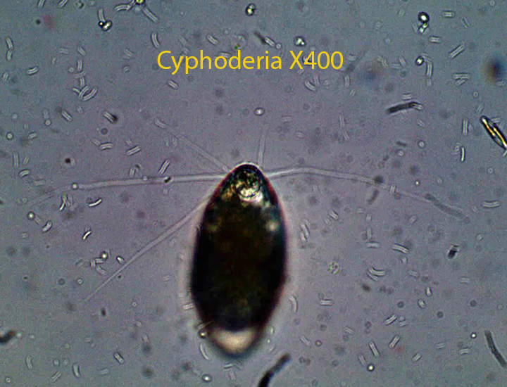
4. Cyphoderia

The smooth, transparent test is rounded posteriorly and curves anteriorly forming a narrow neck, ending with a circular opening. Narrow, filose lobopodia (Filipodia) extend from the opening. Lobopodial expansion and their contraction and extension can move the animal forward, backward, to the right or to the left. Movement is accomplished by extending one pseudopod, attaching it to the substratum and then contracting the entire pseudopod, pulling the animal in that direction.I observed several specimens pull clumps of debris containing small protozoans to the test opening; presumably they were feeding on bacteria and small protozoans.No food vacuoles were found in the clear filipodia suggesting that they are formed inside the test where the filipodia join the cell body.
http://vimeo.com/115670878 (Zottoli) X400
http://vimeo.com/115670879 (Zottoli) X400
http://vimeo.com/115673243 (Zottoli) X400
5. Difflugia (Difflugidae)

Difflugia, about 120 microns long, is housed in a vase-shaped test covered with a few adherent quartz grains. There is a round opening (Aperture) circled by a lip at the anterior end through which the clear pseudopods extend. In one instance, two pseudopodia encircled a detrital mass. Part of the detrital mass containing bacteria and small protozoans was ingested at the base of the tentacles indicating that one role of the tentacles is to move detritus to the cell opening. During this process the pseudopods were clear. Small, round green algal cells (most likely symbiotic) were observed internally along the edge of the cell membrane.
http://vimeo.com/115673244 (Zottoli) X400
http://vimeo.com/115439340 (Zottoli) X400
6. Euglypha (Euglyphidae)

Euglypha, about 230 microns long, is housed in a textured test. The cell surface is covered with flat plates some of which bear needle-like spines. The edge around the oral opening is scalloped. There are no particles adhering to the test. In the first video, thin filliform pseudopodia extend from the single oral opening (Aperture). It is common to see these amoebas with the oral end embedded in detritus containing bacteria and small protozoans, suggesting that they are in feeding mode.
http://vimeo.com/115675913 (Zottoli) X400
http://vimeo.com/115719686 (Zottoli) X600
7. Hartmanella

The cell body, about 20 microns long, is unipodial (One simple tube) with a clear hyaline cap at the tip of the anterior end. A round, clear Contractile Vacuole is present and a posterior uroid bulb is absent. Small, round single cell green algae, possibly symbiotic, found in the cytoplasm.
http://vimeo.com/115675916 (Zottoli) X 400
8. Mayorella (Paramoebidae)

Mayorella spp. (Paramoebidae) is characterized by short, pointed pseudopods that have a broad base. New pseudopods tend to be formed toward the front part of the cell. One contractile vacuole is present. Feeding was not observed.
http://vimeo.com/115711745 (Zottoli) X400
http://vimeo.com/115717508 (Zottoli) X600. A flagellate is trapped in a food vacuole lying inside the cell body.
9 .Saccamoeba (Hartmanellidae)

Saccamoeba, about 30 microns long, has a defined anterior and posterior end. The anterior end always determines the direction of movement and the posterior end follows. The posterior end (Uroid) is of the “Villous Bulb Type”. Small, round green algae were observed in food vacuoles.
http://vimeo.com/115719687 (Zottoli) X400
http://vimeo.com/115719684 (Zottoli) X400
10. Thecamoeba

Thecamoeba, about 50 microns long, is unipodal (one tube). It has a clear anterior hyaline cap and 2-3 longitudinal folds that run the length of the cell body. Diatoms were observed inside several of the specimens.
http://vimeo.com/115719685 (Zottoli) X600
11. Trinema

Trinema, about 30 microns long, has a dorso-ventrally flattened test with overlapping scales. Thin, filose lobopodia (Pseudopods) emerge from the circular, sub-apical test opening. The test is moved from place to place by extension, attachment and contraction of the lobopodia. Often, one pseudopod is extended from the test opening as the animal moves forward. In one instance I observed Trinema placing its pseudopods around a chunk of detritus and directing it into the test through the circular opening.
http://vimeo.com/115722788 (Zottoli) X600
http://vimeo.com/115722787 (Zottoli) X600
Heliozoa:
The cell body of heliozoans consists of an outer cortex that contains the endoplasm and an inner rounded medulla that contains the nucleus and the bases of the needle-like axopods. Each axopod contains a central rod composed of microtubules that is covered with cytoplasm. When prey contact the axopod, they are enclosed in a food vacuole that eventually reaches the cortex.
1. Acanthocystis (Heliozoan)

Acanthocyctis, about 50 microns wide, has numerous parallel-sided arms (long spines or axopodia). Scales can be seen on the external surface of the cell. The shorter spines are forked at the tip.
http://vimeo.com/115439341 (Zottoli) X400
http://vimeo.com/115711743 (Zottoli) X400
2. Actinophyrs (Heliozoan)

Actinophrys, about 100 microns in diameter, is characterized by axopods that taper to a fine pointed tip. The heliozoan has a visible round, centrally located nucleus. Clear food vacuoles are visible along the inside of the cell membrane. A contractile vacuole is visible as a clear bubble next to the cell membrane. They feed for the most part on small protozoans.
http://vimeo.com/115675915 (Zottoli) X400
http://vimeo.com/115711744 (Zottoli) X400
http://vimeo.com/115711741 (Zottoli) X400
3. Actinospherium (Heliozoan)

Actinospherium , with a cell body about 400 microns wide, is characterized by a peripheral layer of large, rounded vacuoles. The multinucleated cells have a number of axopods that taper from the base to the tip, radiating from the cell surface. Both unicellular green flagellates and small rotifers were identified in food vacuoles.
http://vimeo.com/115441419 (Zottoli) X400
http://vimeo.com/115441420 (Zottoli) X400
__________________________________________________________________________________________________________________________________

Leave a comment